Imaging
MRI and MRS
Within the past 30 years, magnetic resonance imaging (MRI) and magnetic resonance spectroscopy (MRS) became the most important method to visualize the entire structure of living organisms. In addition to anatomical imaging, MRI and MRS methods provide valuable insights into the functions of living organisms. MRI and MRS are without any ionizing radiation and without any other type of noteworthy harm to the organisms.
Magnetic resonance imaging (MRI) and spectroscopy (MRS) are noninvasive techniques providing structural and functional insights into living intact organisms with high temporal and spatial resolution.
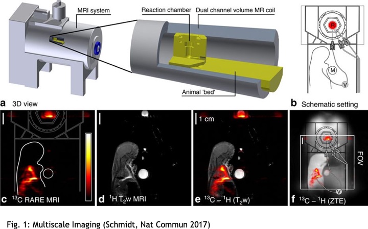
The MOIN CC is equipped with an experimental animal MR system operating at a magnetic field strength of 7 Tesla (ClinScanTM, Bruker BioSpin, Germany). Dedicated coils and a gradient system of 650 mT/m enable our group to analyze any specimens from cell cultures up to animals of the size of rats. In-house developed animal beds allow repeated measurements of anesthetized animals under physiological condition.
As with human magnetic resonance imaging, the study of animals with this method causes no harm to the organisms and can be applied repeatedly to study for instance the course of disease or aspects of aging. Together with the other units of the MOIN CC, truly multimodal imaging is provided for numerous types of biomedical research.